Take Care of Newborn Baby Skin

Take Care of Newborn Baby Skin

Take Care of Newborn Baby Skin
Picture: Strawberry Birth Mark

Diffuse capillary naevi on the face, eyelids, or occiput are common and resolve within a few months.
The “strawberry mark” (Strawberry Angioma) starts as a tiny red spot and grows rapidly for several weeks until it has a raised red appearance with small white areas, suggesting the seeds of a strawberry. Such marks are common in preterm babies. They may occur anywhere on the body but cause no symptoms, except on the eyelids, where they may prevent easy opening of the eyes and need treatment. Strawberry naevi grow, often rapidly, for 3–9 months, but at least 90% resolve spontaneously, either completely or partially. Resolution usually begins at 6–12 months and is complete in half the children by the age of five and in 70% by the age of seven years. In 80% of cases these naevi resolve completely without trace.

The port wine stain is not raised and may be extensive. It does not resolve, but the skin texture remains normal. When the naevus occurs in the distribution of the trigeminal nerve, there may be an associated intracranial vascular anomaly.

Neonatal erythema (erythema “toxicum”) consists of blotchy ill defined areas of bright erythema surrounding white or yellow wheals which may resemble septic spots. It usually appears on the second day of life and in most infants clears within 48 hours. The lesions contain many eosinophils and have no pathological importance. Neonatal erythema is more common in full term infants. By ringing individual lesions with a skin pencil they can be shown to disappear in a few hours, to be replaced by others elsewhere. This contrasts with septic lesions, which appear later and do not resolve so quickly.

Mongolian blue spots are patchy accumulations of pigment, especially over the buttocks and lower back in infants of races with pigmented skins. They are common in babies of African or Mongolian descent, but also occur in Italian and Greek babies. They may be mistaken for bruises and a wrong diagnosis of non-accidental injury made. They become less obvious as the skin darkens.

A midline pit over the spine is most commonly found over the coccyx, where it does not usually communicate with the spinal canal. A midline pit anywhere else along the spine may be connected with an underlying sinus, which may communicate with the spinal canal and requires excision to prevent the entry of bacteria and meningitis.


References:
Bernard Valman. 2002. ABC of the First Year. London: BMJ Books.
Image Source: http://doctorv.ca/cosmetic-services/telangiectasia-broken-facial-blood-vessels/strawberry-hemangioma-strawberry-angioma/  (downloaded Januari 2, 2014 at 22:16 GMT+07)

Umbilical Hernia Symptoms, Causes, Treatment

Umbilical Hernia Symptoms, Causes, Treatment

Umbilical Hernia

An umbilical hernia, usually containing omentum and gut. About 30% of preterm infants who have received mechanical ventilation have an umbilical hernia. No treatment is needed, as the hernia usually disappears spontaneously by the age of three years, although in West Indian infants it may take a further three years.
In contrast to an umbilical hernia the sac of an omphalocoele is covered by peritoneum but incompletely by skin. An omphalocoele is a hernia into the base of the umbilical cord and contains gut and sometimes solid organs like the liver. Immediate transfer to a surgical unit is needed.

Umbilical Hernia Symptoms

Umbilical hernias can usually be seen when your baby is crying, laughing, or straining to use the bathroom. The telltale symptom is a swelling or bulge near the naval area—a symptom not present when your baby is relaxed.
Adults can get umbilical hernias as well. The symptoms will be the same—a swelling or bulge near the navel area. These can be very painful and treatment is usually required.
Symptoms that indicate a more serious situation requiring medical treatment include:
    the baby is in obvious pain
    the baby is vomiting
    the bulge (in both children and adults) is tender, swollen, or discolored

Umbilical Hernia Causes

In a majority of cases, umbilical hernias occur in babies. Babies who are African-American, premature, and of low birth weight are at even higher risk of developing an umbilical hernia. There is no noted difference in occurrence between boys and girls.
An umbilical hernia in adults usually occurs when too much pressure is put on a weak section of the stomach muscles due to:
    being overweight
    frequent pregnancies
    fluid in the abdominal cavity
    stomach surgery


References:
Bernard Valman. 2002. ABC of the First Year. London: BMJ Books.
http://www.healthline.com/health/umbilical-hernia (viewed Januari 2, 2014 at 22:16 GMT+07)
Image Source: http://en.wikipedia.org/wiki/File:Umbilical_Hernia.JPG

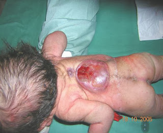
Beware of Myelomeningocele (Spina Bifida) in infants

Beware of Myelomeningocele (Spina Bifida) in infants


Myelomeningocele (Spina Bifida)

A myelomeningocele is a flat or raised neural plaque partly devoid of skin in the midline over the spine due to abnormal development of the spinal cord and associated deficiency of the dorsal laminae and spines of the vertebrae. It is usually found in the lumbar region. The absence of the various coverings that normally protect the cord allows meningitis to occur easily. If there are no active movements in the legs and the anus is patulous, the infant will probably be incontinent of urine and faeces for life and never be able to walk unaided. Thoracic lesions and kyphosis are signs of poor prognosis.
Infants with a good prognosis need urgent treatment, so all affected infants should either be seen by a consultant paediatrician without delay or sent to a special centre, where selection for surgery can be made. About 30% of the infants have surgery as a result of this policy. During the first operation the lesion on the back is covered by skin.
Most of these infants develop progressive hydrocephalus later and those considered suitable for surgery require insertion of a catheter with a valve from a cerebral ventricle to the peritoneal cavity to reduce the cerebrospinal fluid pressure.
Hydrocephalus can be detected by ultrasound examination of the brain. Serial measurements show whether ventricular size is increasing rapidly. In addition, progressive hydrocephalus is confirmed by measuring the circumference of the head at its largest circumference (occipitofrontal) every three days with a disposable paper tape measure, plotting these values on a growth chart, and showing that the head is growing faster than normal.
Raised concentrations of a-fetoprotein are found in the amniotic fluid when the fetus has an open myelomeningocele or anencephaly. In anencephaly there is absence of the cranial vault and most of the brain.
A routine anomaly scan at 18–24 weeks of gestation will detect most neural tube defects and the parents may decide that the pregnancy should be terminated in view of the poor prognosis. Anencephaly is a lethal condition but some infants survive for a few hours or days after birth.


-------------------------------
References: Bernard Valman. 2002. ABC of the First Year. London: BMJ Books. Image source: http://www.flickr.com/photos/laaradj_saadaoui/3461465263/

Psychiatric and social factors of failure to thrive

Psychiatric and social factors of failure to thrive


A physical cause for failure to thrive may be present in a family with psychiatric and social problems and therefore a physical cause must be excluded for all infants. The exclusion of a physical cause is often helpful in persuading the parents to accept that psychiatric or social factors are the main reason for the problem. Features in the history that may suggest this possibility include maternal depression, marital discord, or a disorganised household. Maternal food preferences may result in the exclusion of certain foods, such as cow’s milk, from the diet without reason and without adequate supervision, and may result in a deficient energy intake.

There are more subtle ways in which maternal emotional factors may affect the infant. The mother may be depressed and tense, and this anxiety is transmitted to the child, who does not feed. The mother then reacts by removing the food. In other families the child may be intrinsically less responsive than the average child to food and this may affect the mother’s response to the child at mealtimes. In both these examples the amount of food taken by the child falls to a plateau where the infant appears to be satisfied with the amount given. Another possibility is that the mother keeps to a diet herself as she perceives that she is overweight and also gives a diet to the infant in a similar way.
Some infants are deliberately underfed as part of child abuse. Normal weight gain occurs when fostering has been carried out.

Effective management of infants with this diagnosis requires a team approach involving a health visitor, the family doctor, a child psychiatrist, and paediatrician. Regular advice from a health visitor or dietitian often improves the rate of weight gain.


References:
Bernard Valman. 2002. ABC of the First Year. London: BMJ Books.
Image source: http://emedicine.medscape.com/article/915575-overview
Helicobacter pylori Infection Diagnosis

Helicobacter pylori Infection Diagnosis

Helicobacter pylori Infection Diagnosis

Invasive tests

H pylori can be detected at endoscopy by histology, culture, or urease tests, each with inherent advantages and disadvantages. All these biopsy based methods for detecting H pylori are lible to sampling error because infection is patchy. Up to 14% of infected patients do not have antral infection but have H pylori elsewhere in the stomach, especially if they have gastric atrophy, intestinal metaplasia, or bile reflux. In addition, after partially effective eradication treatment, low levels of infection can easily be missed by endoscopic biopsy, leading to overestimates of the efficacy of eradication treatment and reinfection rates. Proton pump inhibitors affect the pattern of H pylori colonisation of the stomach and compromise the accuracy of antral biopsy. Consensus guidelines therefore recommend that multiple biopsies are taken from the antrum and corpus for histology and for one other method (either culture or urease testing).

Histology—Although H pylori may be recognised on sections stained with haematoxylin and eosin alone, supplementary stains (such as Giemsa, Genta, Gimenez, Warthin-Starry silver, Creosyl violet) are needed to detect low levels of infection and to show the characteristic morphology of H pylori. An important advantage of histology is that, in addition to the historical record provided, sections from biopsies (or even additional sections) can be examined at any time, and that gastritis, atrophy, or intestinal metaplasia can also be assessed. Biopsy specimens from other parts of the stomach can be retained in formalin to be processed only if antral histology is inconclusive.

Culture—Microbiological isolation is the theoretical gold standard for identifying any bacterial infection, but culture of H pylori can be unreliable. Risks of overgrowth or contamination make it the least sensitive method of detection, and it is the least readily available test for use with endoscopy. Although only a few centres routinely offer microbiological isolation of H pylori, the prevalence of multiresistant strains makes it increasing likely that culture and antibiotic sensitivity testing may become a prerequisite for patients with persistent infection after initial or repeated treatment failure.

Urease tests are quick and simple tests for detecting H pylori infection but indicate only the presence or absence of infection. The CLO test and cheaper “home made” urease tests are of similar sensitivity and specificity. However, the sensitivity of urease tests is often higher than that of other biopsy based methods because the entire biopsy specimen is placed in the media, thereby avoiding the additional sampling or processing error associated with histology or culture. The sensitivity of biopsy urease tests seems to be much lower (~60%) in patients with upper gastrointestinal bleeding, but this can be improved by placing multiple biopsy samples into the same test vial.

Non-invasive tests

Serology
H pylori infection elicits a local mucosal and a systemic antibody response. Circulating IgG antibodies to H pylori can be detected by enzyme linked immunosorbent assay (ELISA) antibody or latexagglutination tests. These tests are generally simple, reproducible, inexpensive, and can be done on stored samples. They have been used widely in epidemiological studies, including retrospective studies to determine the prevalence or incidence of infection.

Individuals vary considerably in their antibody responses to H pylori antigens, and no single antigen is recognised by sera from all subjects. The accuracy of serological tests therefore depends on the antigens used in the test, making it essential that H pylori ELISA is locally validated. In elderly people with lifelong infection, underlying atrophic gastritis has been associated with false negative results. Consumption of non-steroidal anti-inflammatory drugs has also been reported to affect the accuracy of ELISAs.

Antibody titres fall only slowly after successful eradication, so serology cannot be used to determine H pylori eradication or to measure reinfection rates. Although titres of IgM antibodies to H pylori fall with age, there are no reliable IgM assays to indicate recent acquisition, which, since this is usually asymptomatic, makes it difficult to identify cases of primary infection and thus establish possible routes of transmission.

An important advantage of serological methods over other tests for H pylori infection has been the development of simple finger prick tests that use a fixed, solid phase assay to detect the presence of H pylori immunoglobulins. These “near patient tests” (NPT) can be performed in primary care and are much simpler than the C-urea breath test, which is the only other test for H pylori that is currently used as a NPT. However, the accuracy of the serological NPT is lower than that reported for standard ELISA tests using the same antigen preparations. These tests are often used to reassure patients, but to date no studies have compared the accuracy, cost effectiveness, and reassurance value of the C-urea breath test with the serological NPT in primary care.

Urea breath test
Non-invasive detection of H pylori by the C-urea breath test is based on the principle that a solution of urea labelled with carbon- will be rapidly hydrolysed by the urease enzyme of H pylori. The resulting CO2 is absorbed across the gastric mucosa and hence, via the systemic circulation, excreted as CO2 in the expired breath. The C-urea breath test detects current infection and is not radioactive. It can be used as a screening test for H pylori, to assess eradication and to detect infection in children. The similar but radioactive C-urea breath test cannot be performed in primary care.

Faecal antigen test
In the stool antigen test a simple sandwich ELISA is used to detect the presence of H pylori antigens shed in the faeces. Studies have reported sensitivities and specificities similar to those of the C-urea breath test ( > 90%), and the technique has the potential to be developed as a near patient test. The main advantage of the test, however, is in large scale epidemiological studies of acquisition of H pylori in children.


References:
- Robert PH Logan. 2002. ABC OF THE UPPER GASTROINTESTINAL TRACT. London: BMJ Books
Helicobacter pylori infection - Definition and Epidemiology

Helicobacter pylori infection - Definition and Epidemiology

Helicobacter pylori infection - Definition and Epidemiology


Helicobacter pylori
is a small, curved, highly motile, Gram negative bacillus that colonises only the mucus layer of the human stomach. Since its discovery in 1984, it has been recognised as the principal cause of peptic ulcer disease and as the main risk factor for the development of gastric cancer. However, most infected people ( > 70%) are asymptomatic. We therefore need to discover how infection is acquired, why ulcers or cancer occur in so few of those infected, and how this subgroup can be identified and treated.

Epidemiology of H pylori infection

H pylori is one of the commonest bacterial pathogens in humans. The prevalence of infection varies but is falling in most developed countries. Seropositivity increases with age and low socioeconomic status. Retrospective seroepidemiological studies have shown a cohort effect consistent with the hypothesis that infection is mainly acquired in early childhood. Until recently, however, it has been difficult to assess accurately the incidence (or route) of infection because of the inaccuracy and cost of detecting (non-invasively) H pylori in young children. Primary acquisition in adults, or reinfection after successful eradication, does occur but is less common, with an annual incidence of 0.3-0.7% in developed countries and 6-14% in developing countries.

How H pylori is usually acquired and its route of transmission are unknown. Since humans are the only known reservoir of infection, it is likely that in developed countries H pylori is picked up from siblings, other children, or parents, predominantly via the gastro-oral route. In developing countries faecal-oral transmission may also occur. Various risk factors are associated with H pylori infection, but the extent to which these are simply markers of childhood socioeconomic deprivation is unclear. H pylori infection is an occupational hazard for gastroenterologists and is associated with performing endoscopy.


References:
- Robert PH Logan. 2002. ABC OF THE UPPER GASTROINTESTINAL TRACT. London: BMJ Books

Heartburn / Gastro-oesophageal reflux disease (GORD) Medical Treatment Guidelines

Heartburn / Gastro-oesophageal reflux disease (GORD) Medical Treatment Guidelines


Aims
For most patients, the aim is acceptable symptom control using the least treatment necessary to achieve this. Therefore, if symptom control is the aim, endoscopy to assess healing of oesophagitis is unnecessary. Indeed, it is now known that, at least for patients treated with proton pump inhibitors, absence of symptoms on treatment equates with healing of oesophagitis. For those with complications, such as stricture or bleeding from oesophagitis, the aim will be long term healing of oesophagitis.
Patients with Barrett’s oesophagus have a risk of between 1 in 50 and 1 in 200 of developing adenocarcinoma of the oesophagus. Many gastroenterologists therefore recommend yearly or biennial endoscopic screening with multiple biopsies to detect dysplasia. Patients with severe dysplasia often have an undetected early cancer and so are offered oesophagectomy. Surveillance of patients with Barrett’s oesophagus to detect severe dysplasia or early cancer is controversial, partly because its benefits have not been established by well designed randomised controlled trials. Clearly a surveillance policy is inappropriate in elderly patients who are unfit for surgery. Endoscopic ablation of the abnormal columnar mucosa in Barrett’s oesophagus by photodynamic laser or thermal methods looks promising and may become standard treatment. It must be combined with high doses of proton pump inhibitors or antirefluxsurgery to prevent continuing acid reflux.

General measures
Patients should be advised to lose weight if overweight. There is no formal evidence to support this assertion, but success (though rarely achieved) may result in improved symptom control. Raising the head of the bed on 15 cm wooden blocks has been shown in a controlled trial to improve symptoms and healing of oesophagitis. There is little evidence that avoidance of specific foods has much effect on the course of the disease, but many patients have already identified and stopped eating foods that produce symptoms before consulting their doctor. Other potentially damaging drug treatment should also be reviewed.
While the benefits associated with these general measures may be unproved, they allow patients to be involved with decision making and may help them avoid over-medicalising their condition.

Antacids and alginates
Antacids are effective for short term relief of symptoms. Although their efficacy is difficult to confirm in controlled trials, many sufferers, particularly those who do not consult a doctor, rely on self medication with antacids. Alginates work by forming a floating viscous raft on top of the gastric contents that provides a physical barrier to prevent reflux. To maximise this effect, they are therefore best taken after meals, otherwise they rapidly empty from the stomach and thus give only transient relief of symptoms by virtue of their antacid content.

Acid suppression therapy
The two major classes of agent available are the H2 receptor antagonists and the proton pump inhibitors. There is little doubt that proton pump inhibitors are more rapidly and completely effective for both relieving symptoms and healing oesophagitis, regardless of disease severity. Because of this, a cost effectiveness argument has been made in favour of proton pump inhibitors as first choice treatments in all cases. However, the data on which these calculations have been made have generally come from hospital based clinical trials and may not be applicable to general practice.
Many patients in primary care may achieve good and lasting symptom relief from short intermittent courses of H2 receptor antagonists at standard doses (such as ranitidine 150 mg twice daily or cimetidine 400 mg twice daily). For patients with severe or refractory oesophagitis, particularly those with complications such as stricture, proton pump inhibitors are the drugs of choice. The optimal daily dose for most patients is omeprazole 20 mg , lansoprazole 30 mg, pantoprazole 40 mg, or rabeprazole 20 mg, but higher doses may give additional clinical benefit in patients with resistant oesophagitis. For most patients, there is no clinical advantage in choosing one proton pump inhibitor over another.

Motility modifying drugs

These include metoclopramide and domperidone. Although both relieve symptoms of heartburn to a degree similar to H2 receptor antagonists, they do not heal oesophagitis. In addition, metoclopramide has a relatively high incidence of side effects on the central nervous system. However, these drugs may be useful, particularly in patients with other dyspeptic symptoms such as nausea or early satiety.

Maintenance treatment
Only proton pump inhibitors, at standard or half standard doses, have been shown to be effective agents for maintenance of remission in those who require it. Indications for maintenance treatment include
  •  Severe oesophagitis, especially presenting with complications (such as stricture, bleeding, peptic ulcers)
  •  Barrett’s oesophagus (although there is no evidence that continuous treatment prevents evolution to cancer)
  •  Symptoms (typical or atypical) relapsing as soon as treatment is stopped.

Surgery
Laparoscopic anti-refluxsurgery seems to be as successful as conventional surgery in controlling refluxin the short term without the disadvantages of a long hospital stay or convalescence. It has become an increasingly popular option for patients requiring long term medical treatment. The results from a randomised controlled trial comparing surgery with maintenance drug treatment are awaited.


References:
- Robert PH Logan. 2002. ABC OF THE UPPER GASTROINTESTINAL TRACT. London: BMJ Books.
- Image Source: http://www.webmd.com/heartburn-gerd/guide/complications-untreated-gerd
Copyright © 2013 Family Medical Center - Dami Tripel Template Level 2 by Ardi Bloggerstranger . All rights reserved.
Valid HTML5 by Ardi Bloggerstranger