Showing posts with label Infants. Show all posts

BreastFeeding Benefits

BreastFeeding Benefits


Breast milk is universally recognized as the preferred source of infant nutrition, and the nutritional advantages of breast milk have been well documented. Colos-trum, the first milk produced after delivery, provides an initial dose of enzymes that promote gut maturation, facilitate digestion and stimulate passage of meco-nium. Colostrum is also high in protein, primarily because of high levels of im-munoglobulins and secretory IgA. The protein in human milk is ideal not only for absorption, but also for utilization, especially by the rapidly developing infant brain. Human milk also contains predominantly polyunsaturated fats with stable amounts of cholesterol, an important constituent of brain and nerve tissue.

Human milk also protects against infection by providing cellular immunity through macrophages and humoral factors, such as antibodies. Numerous studies have verified that breast-fed infants have a lower incidence of bacterial and viral illnesses than bottle-fed infants. This low incidence is of particular clinical significance in developing nations? Ongoing research suggests that breast feeding may provide immuno-logic protection against diabetes mellitus, cancer and lymphoma. Finally, breast feeding has been found to provide protection from allergic diseases, including eczema, asthma and allergic rhinitis. This protection is most likely the result of breast milk decreasing intestinal permeability to large, allergenic molecules.

Recognizing these as well as other advantages, the American Academy of Family Physicians (AAFP) and the American Academy of Pediatrics (AAP) have identified breast milk as the preferred source of infant nutrition. In addition, the U.S. Public Health Service (USPHS) has established a national goal that, by the turn of the century, 75 percent of new mothers will be breast-feeding at the time of hospital discharge. Despite an emphasis on breast feeding by both private and government organizations, only 54 percent of U.S. mothers initiate breast feeding, and fewer than half of these mothers continue nursing for at least six months. Clearly, all health care providers should actively promote breast feeding if the goal set by the USPHS is to be accomplished.

To successfully promote breast feeding, family physicians should consider the influence of marketing campaigns aimed at expectant and new mothers by the manufacturers of infant formulas. Historically, their dogged marketing efforts have included the distribution of free cases of infant formula to expectant mothers, as well as the inclusion of formula samples in commercial hospital discharge packs designed for breast-fed infants. Physicians must work proactively to weigh the risks and benefits of promotional materials and develop appropriate policies governing their distribution in their hospitals or academic institutions.


image source: http://healthynaturalbaby.org/breastfeeding-shown-to-reduce-crib-death-in-infants-by-50/

Take Care of Newborn Baby Skin

Take Care of Newborn Baby Skin

Take Care of Newborn Baby Skin
Picture: Strawberry Birth Mark

Diffuse capillary naevi on the face, eyelids, or occiput are common and resolve within a few months.
The “strawberry mark” (Strawberry Angioma) starts as a tiny red spot and grows rapidly for several weeks until it has a raised red appearance with small white areas, suggesting the seeds of a strawberry. Such marks are common in preterm babies. They may occur anywhere on the body but cause no symptoms, except on the eyelids, where they may prevent easy opening of the eyes and need treatment. Strawberry naevi grow, often rapidly, for 3–9 months, but at least 90% resolve spontaneously, either completely or partially. Resolution usually begins at 6–12 months and is complete in half the children by the age of five and in 70% by the age of seven years. In 80% of cases these naevi resolve completely without trace.

The port wine stain is not raised and may be extensive. It does not resolve, but the skin texture remains normal. When the naevus occurs in the distribution of the trigeminal nerve, there may be an associated intracranial vascular anomaly.

Neonatal erythema (erythema “toxicum”) consists of blotchy ill defined areas of bright erythema surrounding white or yellow wheals which may resemble septic spots. It usually appears on the second day of life and in most infants clears within 48 hours. The lesions contain many eosinophils and have no pathological importance. Neonatal erythema is more common in full term infants. By ringing individual lesions with a skin pencil they can be shown to disappear in a few hours, to be replaced by others elsewhere. This contrasts with septic lesions, which appear later and do not resolve so quickly.

Mongolian blue spots are patchy accumulations of pigment, especially over the buttocks and lower back in infants of races with pigmented skins. They are common in babies of African or Mongolian descent, but also occur in Italian and Greek babies. They may be mistaken for bruises and a wrong diagnosis of non-accidental injury made. They become less obvious as the skin darkens.

A midline pit over the spine is most commonly found over the coccyx, where it does not usually communicate with the spinal canal. A midline pit anywhere else along the spine may be connected with an underlying sinus, which may communicate with the spinal canal and requires excision to prevent the entry of bacteria and meningitis.


References:
Bernard Valman. 2002. ABC of the First Year. London: BMJ Books.
Image Source: http://doctorv.ca/cosmetic-services/telangiectasia-broken-facial-blood-vessels/strawberry-hemangioma-strawberry-angioma/  (downloaded Januari 2, 2014 at 22:16 GMT+07)

Umbilical Hernia Symptoms, Causes, Treatment

Umbilical Hernia Symptoms, Causes, Treatment

Umbilical Hernia

An umbilical hernia, usually containing omentum and gut. About 30% of preterm infants who have received mechanical ventilation have an umbilical hernia. No treatment is needed, as the hernia usually disappears spontaneously by the age of three years, although in West Indian infants it may take a further three years.
In contrast to an umbilical hernia the sac of an omphalocoele is covered by peritoneum but incompletely by skin. An omphalocoele is a hernia into the base of the umbilical cord and contains gut and sometimes solid organs like the liver. Immediate transfer to a surgical unit is needed.

Umbilical Hernia Symptoms

Umbilical hernias can usually be seen when your baby is crying, laughing, or straining to use the bathroom. The telltale symptom is a swelling or bulge near the naval area—a symptom not present when your baby is relaxed.
Adults can get umbilical hernias as well. The symptoms will be the same—a swelling or bulge near the navel area. These can be very painful and treatment is usually required.
Symptoms that indicate a more serious situation requiring medical treatment include:
    the baby is in obvious pain
    the baby is vomiting
    the bulge (in both children and adults) is tender, swollen, or discolored

Umbilical Hernia Causes

In a majority of cases, umbilical hernias occur in babies. Babies who are African-American, premature, and of low birth weight are at even higher risk of developing an umbilical hernia. There is no noted difference in occurrence between boys and girls.
An umbilical hernia in adults usually occurs when too much pressure is put on a weak section of the stomach muscles due to:
    being overweight
    frequent pregnancies
    fluid in the abdominal cavity
    stomach surgery


References:
Bernard Valman. 2002. ABC of the First Year. London: BMJ Books.
http://www.healthline.com/health/umbilical-hernia (viewed Januari 2, 2014 at 22:16 GMT+07)
Image Source: http://en.wikipedia.org/wiki/File:Umbilical_Hernia.JPG

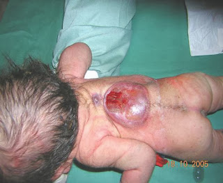
Beware of Myelomeningocele (Spina Bifida) in infants

Beware of Myelomeningocele (Spina Bifida) in infants


Myelomeningocele (Spina Bifida)

A myelomeningocele is a flat or raised neural plaque partly devoid of skin in the midline over the spine due to abnormal development of the spinal cord and associated deficiency of the dorsal laminae and spines of the vertebrae. It is usually found in the lumbar region. The absence of the various coverings that normally protect the cord allows meningitis to occur easily. If there are no active movements in the legs and the anus is patulous, the infant will probably be incontinent of urine and faeces for life and never be able to walk unaided. Thoracic lesions and kyphosis are signs of poor prognosis.
Infants with a good prognosis need urgent treatment, so all affected infants should either be seen by a consultant paediatrician without delay or sent to a special centre, where selection for surgery can be made. About 30% of the infants have surgery as a result of this policy. During the first operation the lesion on the back is covered by skin.
Most of these infants develop progressive hydrocephalus later and those considered suitable for surgery require insertion of a catheter with a valve from a cerebral ventricle to the peritoneal cavity to reduce the cerebrospinal fluid pressure.
Hydrocephalus can be detected by ultrasound examination of the brain. Serial measurements show whether ventricular size is increasing rapidly. In addition, progressive hydrocephalus is confirmed by measuring the circumference of the head at its largest circumference (occipitofrontal) every three days with a disposable paper tape measure, plotting these values on a growth chart, and showing that the head is growing faster than normal.
Raised concentrations of a-fetoprotein are found in the amniotic fluid when the fetus has an open myelomeningocele or anencephaly. In anencephaly there is absence of the cranial vault and most of the brain.
A routine anomaly scan at 18–24 weeks of gestation will detect most neural tube defects and the parents may decide that the pregnancy should be terminated in view of the poor prognosis. Anencephaly is a lethal condition but some infants survive for a few hours or days after birth.


-------------------------------
References: Bernard Valman. 2002. ABC of the First Year. London: BMJ Books. Image source: http://www.flickr.com/photos/laaradj_saadaoui/3461465263/
Copyright © 2013 Family Medical Center - Dami Tripel Template Level 2 by Ardi Bloggerstranger . All rights reserved.
Valid HTML5 by Ardi Bloggerstranger